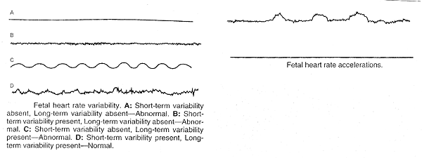
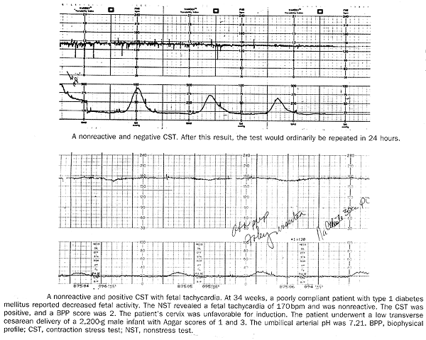
Antepartum Fetal SurveillanceWHEC Practice Bulletin and Clinical Management Guidelines for healthcare providers. Educational grant provided by Women's Health and Education Center (WHEC).The goal of antepartum fetal surveillance is to prevent fetal death. Several antepartum fetal surveillance techniques or tests are in use. These include fetal movement assessment, non-stress test (NST), contraction stress test (CST), biophysical profile (BPP), and umbilical artery Doppler velocimetry. Antepartum fetal surveillance techniques are now routinely used to assess the risk of fetal death in pregnancies complicated by preexisting maternal conditions, as well as those in which complications have developed. Identification of suspected fetal compromise provides the opportunity to intervene before progressive metabolic acidosis can lead to fetal death. However, acute or catastrophic changes in fetal status, such as those that can occur with abruption placentae or an umbilical cord accident, are generally not predicted by tests of fetal well-being. Finally, neither the degree nor the duration of intrauterine hypoxia and academia necessary to adversely affect short and long term neonatal outcome has been established with any precision. The purpose of this document is to review the current indications for and techniques of antepartum fetal surveillance and outline management guidelines for antepartum fetal surveillance, consistent with the best contemporary scientific evidence. Identification of suspected fetal compromise provides the opportunity to intervene before progressive metabolic acidosis can lead to fetal death. In both animals and humans, fetal heart rate pattern, level of activity, and degree of muscular tone are sensitive to hypoxemia and academia. Recent, normal antepartum fetal test results should not preclude the use of intrapartum fetal monitoring. Indications for Antepartum Fetal Surveillance:In general, antepartum fetal surveillance has been employed in pregnancies in which the risk of antepartum fetal demise is increased. Some of the conditions under which testing may be appropriate include the following:- Maternal Conditions:
- Diabetes Mellitus
- Chronic Renal Failure
- Congenital Heart Disease
- Rheumatic Heart Disease
- Thyroid Diseases
- Hemoglobinopathies (hemoglobin SS, SC, Thalassemia)
- Hypertensive disorders
- Pregnancy-related Conditions:
- Pregnancy-induced hypertension
- Decreased fetal movement
- Oligohydramnios
- Polyhydramnios
- Intrauterine growth restriction
- Post-term Pregnancy
- Isoimmunization
- Previous fetal demise
- Multiple gestations
Fetal Movement Assessment:Although several counting protocols have been employed, neither the optimal number of movements nor the ideal duration for counting movements has been defined. Thus numerous protocols for fetal movement assessment by the mother ("kick count") have been reported and appear to be acceptable. In one approach, the woman lies on her side and counts distinct fetal movements. Perception of 10 distinct movements in a period of up to 2 hours is considered reassuring. Once 10 movements have been perceived, the count may be discontinued. In another approach, women are instructed to count fetal movements for 1 hour three times per week. The count is considered reassuring if it equals or exceeds the woman's previously established baseline count. In the absence of a reassuring count, further fetal assessment is recommended. Non-stress Test (NST):The NST is based on the premise that the heart rate of the fetus that is not acidotic or neurologically depressed will temporarily accelerate with fetal movement. Heart rate reactivity is thought to be a good indicator of normal fetal autonomic function. Loss of reactivity is associated most commonly with a fetal sleep cycle but may result from any cause of central nervous system depression, including fetal acidosis. With the patient in the lateral tilt position, the fetal heart rate is monitored with an external transducer. The tracing is observed for fetal heart rate accelerations that peak at least 15 beats per minute above the baseline and last 15 seconds from baseline to baseline. It may be necessary to continue the tracing for 40 minutes or longer to take into account the variations of fetal sleep-wake cycle. Non-stress test (NST) results are categorized as reactive or non-reactive. Acoustic stimulation offers the advantage of safely reducing overall testing time without compromising detection of the acidotic fetus. The NST of the non-compromised preterm fetus is frequently non-reactive in about 50% of times, from 24 to 28 weeks of gestation, and about 15% may not be reactive from 28 to 32 weeks of gestation. Fetal heart decelerations during and NST that persist for 1 minute or longer are associated with a markedly increased risk of both cesarean delivery for a non-reassuring fetal heart rate pattern and fetal demise.  Contraction Stress Test (CST):The CST is based on the response of the fetal heart rate to uterine contractions. It relies on the premise that fetal oxygenation will be transiently worsened by uterine contractions. In the suboptimally oxygenated fetus, the resultant intermittent worsening in oxygenation will, in turn, lead to the fetal heart rate pattern of late decelerations. Uterine contractions also may provoke or accentuate a pattern of variable decelerations caused by fetal umbilical cord compression, which in some cases is associated with oligohydramnios. Contraindications to the CST are: premature labor, preterm membrane rupture, history of extensive uterine surgery or classical cesarean section and known placenta previa. With the patient in the lateral recumbent position, the fetal heart rate and uterine contractions are simultaneously recorded with an external fetal monitor. If at least three spontaneous contractions of 40 seconds' duration each or longer are present in a 10 minute period, no uterine stimulation is necessary. If fewer than three contractions of at least 40 seconds' duration occur in 10 minutes, contractions are induced with either nipple stimulation or intravenous administration of dilute oxytocin. The CST is interpreted according to the presence or absence of late fetal heart rate decelerations, which are defined as decelerations that reach their nadir after the peak of the contraction and that usually persist beyond the end of the contraction. The results of the CST are categorized as follows: - Negative: no late or significant variable decelerations.
- Positive: late decelerations following 50% or more of contractions (even if the contraction frequency is fewer than three in 10 minutes).
- Equivocal- suspicious: intermittent late decelerations or significant variable decelerations.
- Equivocal-hyperstimulatory: fetal heart rate decelerations that occur in the presence of contractions more frequent than every 2 minutes or lasting longer than 90 seconds.
- Unsatisfactory: fewer than three contractions in 10 minutes or an uninterpretable tracing.

 Biophysical Profile (BPP):The BPP consists of an NST combined with four observations made by real-time Ultrasonography. It consists of 5 components: - Non-stress test
- Fetal breathing movements (one or more episodes of rhythmic fetal breathing movements of 30 seconds or more within 30 minutes)
- Fetal movement (three or more discrete body or limb movements within 30 minutes)
- Fetal tone (one or more episode of extension of a fetal extremity with return to flexion, or opening or closing of a hand).
- Determination of the amniotic fluid volume (a single vertical pocket of amniotic fluid exceeding 2 cm is considered evidence of adequate amniotic fluid).
Each of five components is assigned a score of either 2 (normal or present as defined previously) or 0 (abnormal, absent or insufficient). A composite score of 8 or 10 is normal, a score of 6 is considered equivocal, and a score of 4 or less is abnormal. Regardless of composite score, in the presence of oligohydramnios (largest vertical pocket of amniotic fluid volume less than 2 cm), further evaluation is warranted. Modified Biophysical Profile:In the late second or third trimester fetus, amniotic fluid reflects fetal urine production. Placental dysfunction may result in diminished fetal renal perfusion, leading to oligohydramnios. Amniotic fluid volume assessment can therefore be used to evaluate long-term uteroplacntal function. This observation fostered the development of what has come to be termed the "modified BPP" as a primary mode of antepartum fetal surveillance. An amniotic fluid index (AFI) is the sum of measurements of the deepest cord-free amniotic fluid pocket in each of the abdominal quadrants. It is an indicator of long-term placental function. An AFI greater than 5 cm generally is considered to represent an adequate volume of amniotic fluid. Thus the modified BPP is considered normal if the NST is reactive and the AFI is more than 5 and abnormal if either the NST is non-reactive or the AFI is 5 or less. Umbilical Artery Doppler Velocimetry:Doppler Ultrasonography is a noninvasive technique used to assess the hemodynamic components of vascular impedance. It is based on the observation that flow velocity waveforms in the umbilical artery of normally growing fetuses differ from those of growth-restricted fetus. In normally growing fetuses high velocity diastolic flow is seen, whereas with restricted intrauterine growth fetuses diminished umbilical artery diastolic flow is noted. In some cases of extreme intrauterine growth restriction, flow is absent or even reversed. Abnormal flow velocity waveforms have been correlated histopathlogically with small artery obliteration in placental tertiary villi and functionally with fetal hypoxia and acidosis. To maximize interpretability, multiple waveforms should be assessed, and wall-filter settings should be set low enough (typically <150 Hz) to avoid masking diastolic flow. Frequency of Testing:How frequently to perform fetal testing depends on several factors, including clinical judgment. When the clinical condition that prompted testing persists, the test should be repeated periodically until delivery to monitor for continued fetal well-being. If the maternal medical condition is stable and CST results are negative, the CST is typically repeated in 1 week. Other tests of fetal well-being (NST, BPP or modified BPP) are typically repeated at weekly intervals, but in the presence of certain high-risk conditions, such as post-term pregnancy, diabetes mellitus, intrauterine growth restriction, or pregnancy induced hypertension, some investigators have performed twice weekly NST, BPP, or modified BPP testing. Any significant deterioration in the maternal medical status requires fetal reevaluation, as does any acute diminution in fetal activity, regardless of the amount of time that has elapsed since the last test. An abnormal fetal test result should always be considered in the context of the overall clinical picture, taking into account the substantial possibility that the test result is falsely positive. Recent, normal antepartum fetal test results should not preclude the use of Intrapartum fetal monitoring. An abnormal NST or modified BPP usually should be further evaluated by either a CST or full BPP. Subsequent management should then be predicated on the result of the CST or BPP, the gestational age, the degree of oligohydramnios, and the maternal condition. In a term pregnancy complicated by oligohydramnios, delivery is often the most appropriate course of action. How reassuring is a normal test result?In most cases, a normal test result is highly reassuring, as reflected in the false-negative rate of antepartum fetal surveillance, defined as the incidence of stillbirth occurring within 1 week of a normal test result. Based on many studies, the negative predictive value of the NST is 99.8%, and greater than 99.9% of the CST, BPP, and modified BPP. The Doppler velocimetry as the primary means of antepartum fetal surveillance has negative predictive value of 100%. The low false-negative rate of antepartum fetal surveillance tests depends on an appropriate response to any significant deterioration in the maternal clinical status, including retesting of the fetal condition. These tests generally do not predict stillbirths related to acute changes in maternal-fetal status, such as those that occur with abruption placentae or an umbilical cord accident. Moreover, recent, antepartum fetal test results should not preclude the use of intrapartum fetal monitoring. Summary:Women with high-risk factors for stillbirth should undergo antepartum fetal surveillance using the NST, CST, BPP, or modified BPP. Initiating testing at 32-34 weeks of gestation is appropriate for most pregnancies at increased risk of stillbirth, although in pregnancies with multiple or particularly worrisome high-risk conditions, testing may be initiated as early as 26-28 weeks of gestation. Umbilical artery Doppler velocimetry has been found to be of benefit only in pregnancies complicated by intrauterine growth restriction. If used in this setting, decisions regarding timing of delivery should be made using a combination of information from the Doppler Ultrasonography and other tests of fetal well-being, along with careful monitoring of maternal status.Suggested Reading:- ACOG Practice Bulletin. Antepartum fetal surveillance. Number 9, October 1999
- Dudley NJ. A systemic review of ultrasound estimation of fetal weight. Ultrasound Obstet Gynecol 2005;25:80-89
- Kontopoulous EV, Vinzileos AM. Condition-specific antepartum fetal testing. Am J Obstet Gynecol 2004;191:1546-1548
- Fretts RC. Etiology and prevention of still births. Am J Obstet Gynecol 2005;193:1923-1925
- Kahn B, Lumey LH, Zybert PA et al. Prospective risk of fetal death in singleton, twin and triplet gestation: implications for practice. Obstet Gynecol 2003;102:685-689
- Martin JA, Hamilton BE, Sutton PD et al. Births: final data for 2002. Natl Vital Stat Rep 2003;52:1-3
- March of Dimes Perinatal Profiles: Health indicators, 1990-1997. New York, The March of Dimes Perinatal Data Center and Planning and Community Service Division, 2000
- Morbidity and Mortality Weekly Report: Trends in infant mortality attributable to birth defects -- United States, 1980-1995. Boston, Massachusetts Medical Society, 2000
- Divion ML, Ferber A. Evidence based antepartum fetal testing. In Prenatal and Neonatal Medicine. New York, The Parthenon Publishing Group, 2000
- Druzin ML, Smith JF, Gabbe SG et al. Antepartum fetal evaluation. In Obstetrics: Normal and Abnormal Pregnancies. 5th edition, Churchill Livigstone, an imprint of Elsevier Inc. 2007
- Stanton C. Steps towards achieving skilled attendance at birth. Bulletin of the World Health Organization 2008;86:242
©
Women's Health and Education Center (WHEC)
|