


Cervical Cancer: Early Detection and Prevention
WHEC Practice Bulletin and Clinical Management Guidelines for healthcare providers. Educational grant provided by Women's Health and Education Center (WHEC).
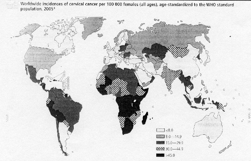
Worldwide, cervical cancer is the second most common malignancy in women and a major cause of morbidity and mortality. Cervical cancer is gender-specific disease that disproportionately affects women in the lowest socioeconomic classes throughout the world. In 2004, the 57th World Health Assembly adopted World Health Organization's global reproductive health strategy, which identified five priority areas including "combating sexually transmitted infections"; the strategy also specifically addressed cervical cancer prevention. Screening programs have successfully reduced disease rates in developed countries that support cytology-based services; these services are too complex for most developing countries to implement. More than 80% of the estimated 500,000 incident cases annually and more than 90% of the 257,000 deaths caused by cervical cancer occur in developing countries. This disparity is due in large part to the fact that a majority of women in these countries have never been screened for cervical cancer. Human papillomavirus (HPV) is a sexually transmitted infection, recognized as the necessary cause of 99% of all cervical cancers. Since last year, it has become possible to vaccinate against the human papillomavirus (HPV) that causes most cases of cervical cancer, but countries face tough decisions before making the vaccine widely available. There are challenges for countries in terms of cost and so on. Meanwhile, the World Health Organization has been developing information that countries can use to formulate their policies on HPV vaccination.
The purpose of this document is to highlight early detection of cervical cancer and prevention. Many important advances have also taken place in the diagnosis and treatment of cervical cancer. This review also defines the strategies for diagnosis and management of abnormal cervical cytology and histology. These strategies reflect new information concerning the natural history of cervical carcinogenesis and the performance of screening and diagnostic tests. The most important component in the management of cervical cancer will always be primary prevention. Sophisticated new tests for the detection of HPV that hold great promise for improved screening abnormal screening for cervical cancer precursors and invasive cancer and for the triage of abnormal cervical cytology also have been developed. Understanding the immunology of HPV has allowed the development of new and more effective treatment modalities of HPV infection and the preliminary development of primary prevention modalities, including HPV vaccines.
Abbreviations:
HPV - human Papillomavirus
ASC-US - atypical squamous cells of undetermined significance
LSIL - low-grade squamous intraepithelial lesion
HSIL - high-grade squamous intraepithelial lesion
CIN - cervical intraepithelial neoplasia
AGC - atypical glandular cells
AIS - adenocarcinoma in situ
Incidence / Burden of Disease:
A resolution on cancer prevention and control was adopted by WHO's Member States, and a new vision and strategy for global immunization that aims to ensure equal access to immunization for every child, adolescent and adult was endorsed during the 58th World Health Assembly in 2005. With the upcoming introduction of a vaccine to prevent human papillomavirus (HPV) infection, a comprehensive approach to preventing cervical cancer - which incorporates vaccination, screening and early treatment - opens up new opportunities for strengthening reproductive health services and building interdisciplinary links (1). Adolescent health programs are developing user-friendly services that aim to provide counseling on sexual health that focuses on the prevention of pregnancy and sexually transmitted infections including HIV. The presence of a new intervention, such as an HPV vaccine, could extend the scope of these services and help to integrate other interventions, thereby making them more attractive to young people.
Worldwide incidences of cervical cancer per 100,000 females (all ages), age-standardized to the WHO standard population, 2005
(Source: World Health Organization)
Epidemiology:
The past decade has seen a remarkable increase in the knowledge of the natural history of cervical dysplasia. Although most infections with HPV are not clinically detectible, the most commonly recognized visible HPV-induced lesion of the female lower genital tract is genital or venereal warts (condylomata acuminate). More than 15 years ago, a relationship between HPV infection and cervical cancer was recognized. Since then, important strides in understanding the virus have been made, particularly in the following areas: modes of transmission and risk factors associated with transmission; the oncogenic potential of specific viral types and the mechanism by which they cause cancer; and the spectrum of infection, ranging from asymptomatic carrier states to overt warts, pre-neoplastic lesions, and invasive cancer. HPV is a sexually transmitted infection, recognized as the necessary cause of 99% of all cervical cancers. HPV is highly prevalent, with an estimated 20 million people infected in the United Sates. The incidence of HPV infections is rising, with 6.2 million new infections diagnosed annually. Cumulative prevalence rates are as high as 82%, underscoring the fact that all sexually active individuals are at high risk of acquiring HPV infection and are subject to developing HPV-associated diseases. Risk factors for disease include multiple partners, high parity (five or more pregnancies), smoking, impaired cellular immunity, and a young age at the instigation of sexual relationships (2). Every year in the United States, between 2 and 3 million women are identified with cervical cytology showing ASCUS, and 1.25 million women are identified with low-grade squamous intraepithelial lesions (LSIL).
Human Papillomavirus (HPV) is a small DNA virus with a genome of approximately 8,000 base pairs. More than 100 putative HPVs have been described; approximately 40 HPV types can be found in the cervico-vaginal area. The development of cervical carcinoma is restricted to a subset of viruses that increase the risk of cervical cancer in infected women (high-risk), of which HPV 16 and HPV 18 together account for approximately two thirds of observed cervical cancer cases. The ability of HPV infections to progress to malignancies is caused in large part by the biochemical activities of E6 and E7, the primary oncoproteins encoded by HPV genomes. Although other oncoproteins, such as E4 and E5, are thought to complement their activity, E6 and E7 from high-risk types manipulate cell cycle regulators, induce chromosomal abnormalities, and block apoptosis. HPV targets the basal cells in the stratified squamous epithelium and the metaplastic cells in the stratified squamous epithelium and metaplastic cells at the squamocolumnar junction of the cervix. Additionally, HPV may infect the glandular epithelium of the endocervix, resulting in glandular neoplasms, such as adenocarcinoma in situ or invasive adenocarcinoma (3). Steps that occur from initial infection leading to the development of cancer include overcoming host immune resistance, possible integration of HPV DNA into the host genome, and accumulation of additional mutations within the infected host cell. HPV must be persistent within the host epithelial cells as a preliminary step forward toward advanced neoplastic changes.
The 2001 Bethesda System Categorizing of Epithelial Cell Abnormalities (4):
Squamous cell
- Atypical squamous cells (ASC)
- Of undetermined significance (ASC-US)
- Cannot exclude HSIL (ASC-H)
- Low-grade squamous intraepithelial lesions (LSIL)
- Encompassing human papillomavirus (HPV), mild dysplasia, and cervical intraepithelial neoplasia (CIN) 1
- High-grade squamous intraepithelial lesions (HSIL)
- Encompassing moderate and severe dysplasia, carcinoma in situ, CIN 2, and CIN 3
- Squamous cell carcinoma
Glandular cell
- Atypical glandular cells (AGC) (specify endocervical, endometrial, or not otherwise specified)
- Atypical glandular cells, favor neoplastic (specify endocervical or not otherwise specified)
- Endocervical adenocarcinoma in situ (AIS)
- Adenocarcinoma
Natural History of HPV and CIN:
Most women infected by HPV are asymptomatic. The virus is detected by an abnormal Pap test result, HPV test result, or the presence of clinically evident genital warts, and most likely will resolve without treatment. Carriage of HPV DNA is extremely common in the general population, reported in one study to occur at least once over a 3-year period in 60% of young women (5). Extrapolating from these reported acquisition rates, the lifetime cumulative risk is at least 80%. The vast majority of women clear the virus or suppress it to levels not associated with CIN 2/3+, and for most women this occurs promptly. The duration of HPV positivity is shorter and the likelihood of clearance is higher in younger women. The presence of HPV is a marker for the risk of diagnosis of CIN 2/3+; only 1 in 10 to 1 in 30 HPV infections are associated with abnormal cervical cytology results, with an even smaller proportion associated with CIN 2/3+. Among women with negative cytology and a positive HPV test result, only 15% will have abnormal cytology results within 5 years. However, HPV is necessary for the development and maintenance of CIN 3 (6). In longitudinal studies, the time interval required for 50% of prevalent cases to become HPV DNA negative for the HPV type detected at enrollment was 4.8 months for non-oncogenic types and 8.1 months for oncogenic types. However, this is not a true measure of the duration of infections because it is unknown how long the women had been infected by the time they were found to be positive at enrollment. In another longitudinal study, the duration of infection was determined for incident infection. The median duration of HPV DNA was 8 months.
From a clinical perspective, it is important to distinguish which intraepithelial neoplasia will progress to invasive cancer if left untreated. However, the diagnostic categories currently available have only modest predictive value, and that value decreases as the lesions become less severe. Cancer precursors include CIN 3, AIS, and to a lesser extent CIN 2. The likelihood of progression to cancer is higher and the time to progression is shorter as the grade of dysplasia increases. Although expression of the presence of HPV as CIN can occur within months of viral acquisition, the time course from CIN 3 to invasive cancer averages between 8.1 and 2.6 years. The leisurely pace of these changes in immuno-competent women means that accurate estimates of progression risk require long follow-up times. Perhaps more relevant for clinical practice are estimates of regression to normal. The goals of cervical screening are not to prevent CIN but to prevent and detect early invasive cervical cancer and to reduce mortality. The detection of CIN 2 or CIN 3 is not a screening failure but rather the goal of the screening system. In natural history studies of adolescents with newly acquired HPV infection, the average length of detectable HPV is 13 months. In most adolescent patients with an intact immune system, an HPV infection will resolve within 24 months. Further evidence that the HPV infection will resolve without treatment comes from the high rates of resolution of CIN 1 and CIN 2, 70% and 50% respectively.
The Pap Smear
HPV DNA Testing:
The strong correlation between infection with high-risk types of HPV and LSIL, HSIL, and cervical cancer suggests that HPV DNA testing would be a useful tool for the management of women with abnormal Pap test results, especially in the case of those with equivocal test results. In the case of an equivocal Pap test result, HPV DNA testing can help determine whether the individual should be referred for colposcopic assessment. In 2001, the National Cancer Institute completed a large multicenter randomized trial called the Atypical Squamous Cell of Undermined Significance/Low Grade Squamous Intraepithelial Lesion Triage Study (ALTS). The study compared immediate colposcopy, HPV DNA testing by Hybrid Capture 2, and repeat cytology in the management of women with ASC-US and LSIL cytology. The study showed that 83% of women with LSIL cytology tested positive for high-risk HPV DNA, limiting the usefulness and the cost-effectiveness of HPV testing in differentiating which women with LSIL cytology should undergo colposcopy. In women with ASC-US cervical cytology screening results, ALTS found that triage using liquid-based cytology alone had a sensitivity of 85% for CIN 2 and CIN 3 lesions while referring 59% of women to colposcopy following the first repeat cervical cytology screening and 67% following the second repeat cervical cytology screening. Human papillomavirus testing detected more than 90% of CIN 2 and CIN 3 while referring only 56% of the patients to colposcopy. Both HPV testing and repeat cytology referred more women younger than 30 years to colposcopy than older women, but HPV testing was significantly more sensitive for CIN 2 and CIN 3 at all ages (7). Testing for HPV DNA has clinical relevance for predicting the risk of the presence of CIN 2/3+ and the risk of the development of CIN 2/3+. The American Society for Colposcopy and Cervical Pathology (ASCCP) consensus conference recommended that HPV DNA testing could be used as a test of cure for women with CIN 2 or CIN 3 at least 6 months following excision or ablation of the transformation zone. The women with high-risk HPV would be referred for colposcopy.
Management of Abnormal Cervical Cytology and Histology:
In Adolescents: the management of abnormal cervical cytology in adolescents differs from that for the adult population in many cases. Certain characteristics of adolescents may warrant special management considerations. It is important to avoid aggressive management of benign lesions in adolescents because most cervical intraepithelial neoplasia grades 1 and 2 regress. Surgical excision or destruction of cervical tissue in a nulliparous adolescent may be detrimental to future fertility and cervical competency. A compliant, health-conscious adolescent may be adequately served with observation in many situations. The following recommendations are unique to the adolescent population and address the clinical situations that can be managed by cytologic follow-up, HPV testing, colposcopy, or a combination of these approaches. A positive HPV test result refers to the presence of high-risk HPV DNA as determined by Hybrid Capture II. Testing for low-risk HPV types has no role in cervical cancer prevention. Adolescents with ASC-US who have an HPV test result negative for high-risk HPV DNA should have a Pap test in 12 months. LSIL test results can be monitored by repeat cytology at 6-month intervals or by a high-risk HPV test in 12 months. These individuals should undergo colposcopy for any cytologic abnormality or the persistence of HPV infection at 1 year. Immediate colposcopy is an acceptable alternative for adolescents with LSIL (8). High-grade squamous intraepithelial lesions (HSIL) are a significant cytologic abnormality that requires colposcopic evaluation because of a much higher rate of histologically confirmed CIN 2, CIN 3, or cervical cancer. The specific method of follow-up should be individualized by the health care professional. A reasonable approach to the follow-up could be either cytology or colposcopy at 4-6-month intervals.
Normal cytology with positive HPV testing: the use of high-risk HPV testing as an adjunct to cervical cytology in women aged 30 years and older is recently introduced. Women with a normal cytology result who test positive for HPV on routine screening have a risk of CIN 2/3 of approximately 4%, which is lower than the risk for women with ASC cytology results. For this reason, colposcopy is not recommended as further testing after a single HPV-positive, cytology-negative result. Current expert consensus recommendations include a repeat cytology and HPV test in 6-12 months to allow for resolution of transient HPV carriage and a colposcopy only if test results remain abnormal (HPV-positive or ASC or higher-grade cytology results) (9).
Atypical squamous cells (ASC) cervical cytology: options for evaluation include immediate colposcopy, triage to colposcopy by HPV DNA testing, or repeat cytology test at 6 and 12 months. Aggressive investigation should be avoided because ASC diagnosis is poorly reproducible between observers, the risk of cancer is very low (0.1-0.2%), and the risk of CIN 2/3+ for any individual patient is also low (6.4-11.9%). Testing for oncogenic HPV and referral to colposcopy only for those women who test positive has the advantage of prompt diagnosis and the ability to reassure 44-69% of patients without colposcopy that their risk of a significant lesion is very low. If HPV testing is elected, those women whose test results are HPV-positive have a 15-27% chance of having a CIN 2/3+ and should be referred for colposcopy. Those women who test negative for HPV can be reassured that their risk of harboring CIN 2/3+ is less than 2%, and they can be scheduled for repeat cytology testing in 1 year. The exception to this recommendation for HPV follow-up is the adolescent, for whom the risk of invasive cancer approaches zero and the likelihood of HPV clearance is very high. At least two consecutive follow-up cytology tests, with referral to colposcopy for an ASC or higher-grade result, are required to reach the sensitivity of a single HPV test for the detection of CIN 2/3+. Although follow-up cytology tests may allow some women to avoid colposcopy, waiting 6-12 months for a definitive diagnosis can create anxiety and possible delay in diagnosis of cancer. In addition, the rate of loss of follow-up is substantial, with 15-25% reported in the research setting and 54-81% non-compliance in clinical practice settings (10).
Low-grade squamous intraepithelial lesion (LSIL) cervical cytology: a cytology result of LSIL is the second most common abnormal cytology result and is more frequent in younger populations with larger number of recent partners. An LSIL diagnosis is associated with a positive test result for high-risk HPV in most women (83% of the women with LSIL cytology in ALTS); therefore HPV testing is of limited value in triage to colposcopy. The risk of CIN 2/3+ at initial colposcopy following and LSIL result is between 15% and 30% in most studies. Therefore, colposcopy is recommended for evaluation of LSIL. The cytology result "atypical squamous cell - can not exclude HSIL" (ASC-H) is intended to include 5-10% of ASC cases overall. Findings of CIN 2/3+ in 24-94% of patients with ASC-H cytology results suggest that colposcopy is an appropriate initial diagnostic intervention. This terminology is used to alert clinicians that the risk of CIN 3 or cancer exceeds that of ASC findings but lacks the certainty required for the patient to be given and HSIL interpretation, which could prompt an excision if colposcopic evaluation was negative (11). In women aged 30 years or older with ASC-H cytology results, HPV-positive test results decrease dramatically, and triage to colposcopy using HPV testing may be considered as with other ASC findings. Otherwise, women with ASC-H should be cared in a fashion similar to women with LSIL; follow-up of a colposcopy result of CIN 1 or normal should include either cytology tests at 6 and 12 months or an HPV DNA test at 12 months, rather than excision.
High-grade squamous intraepithelial lesion (HSIL) cervical cytology: among women with HSIL cytology results, CIN 2 or CIN 3 have been reported in 70% or more, and 1-2% harbor invasive cancer (12). Given the level of risk, colposcopy and biopsy of visible lesions are recommended. Endocervical assessment should be performed in the non-pregnant patient, and the entire vagina should be examined, especially when a lesion corresponding to the cytology result is not found. An alternative "see and treat" management plan may be used in this patient population in the event that a lesion consistent with CIN 2 or CIN 3 is seen. In women who have such a lesion and are not pregnant, LEEP may be performed at the same visit as the colposcopy. In these cases, the cervical biopsy is omitted and an endocervical assessment after the LEEP may be considered.
Atypical glandular cells (AGC) or Adenocarcinoma in situ (AIS) in cervical cytology: the risk of CIN 2/3+ in women with AGC cytology results is 9-41%, in contrast to 27-96% with AGC favor neoplasia. At least half of AIS and adenocarcinoma results will be accompanied by squamous CIN. An AIS cytology result is associated with a 48-69% risk of histologic AIS and a 38% risk of invasive adenocarcinoma of the cervix (13). Endocervical curettage and colposcopy are both relatively insensitive for AIS and adenocarcinoma, but most women with AGC cytology results do not have significant lesion. The initial evaluation of women with AGC results is dictated by the risk of CIN 2/3+ noted previously, by the possibility that the source of the abnormality may be the endometrium, and by the recognition that the entire endocervix is at risk for AIS, mandating endocervical sampling. As a consequence, colposcopy and endocervical sampling should be included in the initial evaluation of all women with AGC result, except for those with results that specify "atypical endometrial cells". Women with atypical endometrial cells and a normal endometrial sampling should undergo colposcopy and endocervical sampling. Endometrial sampling is indicated in women with atypical endometrial cells and all women with AGC results who are aged 35 years or older, as well as those younger than 35 years with abnormal bleeding, morbid obesity, oligomenorrhea, or clinical evaluation suggesting endometrial cancer.
Human papillomavirus (HPV) vaccines:
A new quadrivalent vaccine, Gardasil, protects against four HPV types (6, 11, 16, 18), which are responsible for 70% of cervical cancers and 90% of genital warts. This prophylactic vaccine is made from non-infectious HPV-like particles (or virus-like particles, VLP); it does not contain thimerosal or mercury. The vaccine is administered through a series of three intramuscular injections over a six-month period (at 0, 2, and 6 months). On June 8, 2006 this vaccine was licensed by the Food and Drug Administration (FDA), becoming the first licensed vaccine developed to prevent cervical cancer and other diseases in females caused by genital HPV infection. On June 29, 2006 the Advisory Committee on Immunization Practices (ACIP) voted to recommend use of the quadrivalent vaccine in females, ages 9 to 26 years of age. Ideally, the vaccine should be administered before onset of sexual activity. However, females who are sexually active also may benefit from vaccination. Females who already have been infected with one or more HPV type would only get protection from the vaccine type(s) they have not acquired. Clinical trials have demonstrated 100% efficacy in preventing cervical pre-cancers caused by the targeted HPV types and nearly 100% efficacy in preventing vulvar and vaginal pre-cancers and genital warts caused by the targeted HPV types among women ages 16 to 26 years, who were naïve to the specific HPV vaccine types. Data do not indicate that the vaccine has any therapeutic effect of HPV infection or HPV-associated disease, including existing Pap test abnormalities or genital warts (14). While it is possible that vaccination of males may offer direct health benefits to males and indirect health benefits to females, there are currently no efficacy data available to support use of HPV vaccine in males. Efficacy studies in males are ongoing.
Editor's Note:
Women with ASC cytology results may undergo immediate colposcopy, triage to colposcopy by high-risk HPV DNA testing, or repeat cytology screening at 6 and 12 months. Triage to colposcopy should occur after positive HPV test results of ASC or higher-grade diagnosis. Women with ASC who test negative for HPV or whose HPV status is unknown and test negative for abnormalities using colposcopy should have a repeat cytology test in 1 year. Most women with ASC who are HPV positive or women with ASC-H, LSIL, or HSIL test results should undergo colposcopy. The recommendation for follow up of untreated CIN 1 includes cytology test at 6 and 12 months with colposcopy for an ASC or higher-grade result, or a single HPV test at 12 months, with colposcopy if the test result is positive. Pregnant women with CIN 2 or CIN 3 may undergo follow-up with colposcopy during each trimester and should be reevaluated with cytology and colposcopy examinations at 6-12 weeks postpartum or thereafter. Treatment of CIN 2 and CIN 3 in pregnancy is not indicated. Women with CIN 2 or CIN 3 should be treated (in absence of pregnancy) with excision or ablation. Management of CIN 2 in adolescents may be individualized.
Approximately half of all cervical cancers occur in women who have never been screened. Therefore, screening is particularly important in women who have never or rarely been screened. HPV vaccine providers should notify vaccinated females that: they will need regular cervical cancer screening as the vaccine will not provide protection against all types of HPV that cause cervical cancer; they should practice protective sexual behaviors (e.g., abstinence, monogamy, limiting their number of sex partners, and using condoms), as the vaccine will not prevent all cases of genital warts - nor will it prevent other sexually transmitted diseases; they may not receive the full benefits of vaccine after they have become sexually active (and may have already acquired a vaccine HPV type) or if they do not complete the full vaccine series.
Resources:
- World Health Organization
Planning and implementing cervical cancer prevention and control programs: A manual for managers - American Society for Colposcopy and Cervical Pathology
CONSENSUS GUIDELINES: Guidelines on Management of Women with Histological Abnormalities - US Food and Drug Administration
Quadrivalent human papillomavirus (types 6, 11, 16, 18) recombinant vaccine, 2006 (product approval information: licensing action). - U. S. National Institutes of Health (NIH)
Cervical Cancer
References:
- Pollack AE, Balkin M, Edouard L et al. Ensuring access to HPV vaccine through integrated services: a reproductive health perspective. Bulletin of the World Health Organization 2007;85:57-63
- Centers for Disease Control and Prevention. Genital HPV Infection - CDC Fact Sheet. Centers for Disease Control and Prevention. 2004
- Scheurer ME, Tortolero-Luna G, Adler-Storthz K. Human Papillomavirus infection: biology, epidemiology, and prevention. International J Gynecology Cancer 2005;15(5):727-746
- ACOG Practice Bulletin. Management of Abnormal Cytology and Histology. Number 66, September 2005.
- Einstein MH, Burk RD. Persistent human papillomavirus infection: definitions and clinical implications. Papillomavirus Rep 2001;12:119-123. (Level III)
- Castle PE, Wacholder S, Sherman ME et al. Absolute risk of a subsequent abnormal pap among oncogenic human papillomavirus DNA-positive, cytologically negative women. Cancer 2002;95:2145-2151. (Level II-2)
- Sherman ME, Schiffman M, Cox JT. Effects of age and human papilloma viral load on colposcopy triage: data from the randomized Atypical Squamous Cells of Undetermined Significance/Low-Grade Squamous Intraepithelial Lesion Triage Study (ALTS). J Natl Center Inst 2002;94:102-107
- ACOG Committee Opinion. Evaluation and management of abnormal cervical cytology and histology in the adolescent. Number 330, April 2006.
- Wright TCJr, Schiffman M, Solomon D et al. Interim guidance for the use of human papillomavirus DNA testing as an adjunct to cervical cytology for screening. Obstet Gynecol 2004;103:304-309. (Level III)
- Kim JJ, Wright TC, Goldie SJ. Cost-effectiveness of alternative triage strategies for atypical squamous cells of undermined significance. JAMA 2002;287:2382-2390. (Level III)
- Guido R, Schiffman M, Solomon D, Burke L. Post-colposcopy management strategies for women referred with low-grade squamous intraepithelial lesions or human papillomavirus DNA-positive atypical squamous cells of undetermined significance: a two-year prospective study. ASCUS LSIL Triage Study (ATLS) Group. Am J Obstet Gynecol 2003;188:1401-1405. (Level II-2)
- Jones BA, Davey DD. Quality management in gynecologic cytology using inter-laboratory comparison. Arch Pathol Lab Med 2000;124:672-681. (Level III)
- Oliveira ER, Derchain SF, Rabelo-Santos SH et al. Detection of high-risk human papillomavirus (HPV) DNA by Hybrid Capture II in women referred due to atypical glandular cells in the primary screening. Diagn Cytopathol 2004;31:19-22. (Level III)
- Villa LL, Costa RL, Petta CA et al. Prophylactic quadrivalent human papillomavirus (types 6, 11, 16, and 18) L1 virus-like particle vaccine in young women; a randomized double-blind placebo-controlled multicenter phase II efficacy trial. Lancet Oncol 2005;6(5):271-278
Published: 24 June 2009
Dedicated to Women's and Children's Well-being and Health Care Worldwide
www.womenshealthsection.com